Articles
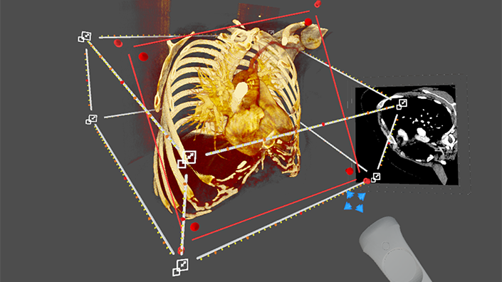
That could soon change, however, thanks to a system developed at King’s College London (KCL) and Evelina London Children’s Hospital. The technology combines multiple scans into a beating, three-dimensional ‘digital double’ of the heart that can be viewed and manipulated in virtual reality (VR).
“The VR really gives you that intuitive ability to ‘pick up’ and move the heart around, they can position it in whatever orientation they want,” said Dr Natasha Stephenson, clinical research fellow at the KCL School of Biomedical Engineering and Imaging Sciences. “It’s really about having that immersion, that intuitiveness, and that understanding of 3D relationships.” The team, which received funding from the British Heart Foundation, hopes the system could improve outcomes for the thousands of patients who undergo surgical or keyhole procedures for congenital heart disease each year.
The program integrates echocardiograms – ultrasound scans of the heart – with computed tomography (CT) and magnetic resonance imaging (MRI), which are typically viewed on a flat screen. 3D models of real medical devices are also being added to the system, so surgeons can try out different approaches.
Against the clock
In the UK, an average of 13 babies are diagnosed with congenital heart disease each day. Depending on the severity of their condition, they might need one or more procedures to help their hearts function normally. No two hearts are the same, but the KCL system shows surgeons exactly what they are going to find, helping them plan ahead and to take the right course of action. The heart is normally stopped during procedures, meaning time is of the essence.
“In the group of patients we focused on, all of their hearts and heart defects were slightly different. You don’t find two people the same,” said lead researcher Professor John Simpson. “Once the heart is stopped, machines take over the work of the heart. But, essentially, the clock is ticking... surgeons want to know exactly what they’re going to find, and exactly what they’re going to do about it.”

The program integrates echocardiograms with CT and MRI scans
The approach aims to prevent any surprises, minimising operating time and making it more predictable. It can also ensure the correct size of medical devices are used, preventing complications. Overall, it could reduce the need for multiple surgeries, leading to better outcomes and experiences for patients and families.
The system is designed to be as practical and as useful as possible. Training takes a matter of minutes, and in-built rendering features help avoid time-intensive segmentation of the initial scans.
Surgeons who trialled a version using just echocardiograms preferred it for understanding the anatomy of patients’ hearts, KCL said, while they also reported increased confidence and improved decision making.
Other benefits
Other procedures that could benefit from a similar approach include orthopaedic surgery, reconstruction of bones and faces, and abdominal surgery, said the researchers. They hope the system could be in regular use within two years, and are working with the KCL quality management team to help it reach national regulatory standards.
Despite the exciting promise of the project, it cannot be a case of ‘VR for VR’s sake’ in such a safety-critical sector. “We have zero interest in introducing VR just to produce another image,” said Simpson. “It just doesn’t fly – it’s got to offer people something more than they currently get.”
Want the best engineering stories delivered straight to your inbox? The Professional Engineering newsletter gives you vital updates on the most cutting-edge engineering and exciting new job opportunities. To sign up, click here.
Content published by Professional Engineering does not necessarily represent the views of the Institution of Mechanical Engineers.